Technologies for
Genomic & Proteomic function
Europe: tel. +32 1658 9045 | fax: +32 1650 9045
More information on our contact page
Products:
Product Brochures:
Cumate Switch Inducible Vectors
Sterol Sensing pGreenFire Reporters
Other Products:
Our Partner:

Precipitate Exosomes from Biofluids
Overview:


Discover microRNA and Protein Biomarkers in Patient Biofluids
Exosomes are 40 –100 nm membrane vesicles secreted by most cell types in vivo and in vitro. Exosomes are found in blood, urine, amniotic fluid, malignant ascite fluids and contain distinct subsets of microRNAs depending upon the tumor from which they are secreted. SBI's ExoQuick exosome precipitation reagent makes microRNA and protein biomarker discoveries simple, reliable and quantitative. Enrich for circulating exosomal microRNAs with ExoQuick and accurately profile them using SBI’s QuantiMir qPCR arrays.
* No time-consuming ultracentrifugation
* Less expensive than costly Antibodies and beads
* No complicated syringes required
* Compatible with biofluid from any species
* More effective than any other method
* Isolate intact exosomes for functional studies
* Use as little as 100 µl of serum or biofluid
Isolate Exosomes from Culture Media and Urine with ExoQuick-TC
Discover microRNA and Protein Biomarkers in Patient Biofluids
Exosomes are 40 - 100 nm membrane vesicles secreted by most cell types in vivo and in vitro. Exosomes are found in blood, urine, amniotic fluid, malignant ascite fluids and contain distinct subsets of microRNAs depending upon the tumor from which they are secreted. SBI's ExoQuick exosome precipitation reagent makes microRNA and protein biomarker discoveries simple, reliable and quantitative. Enrich for circulating exosomal microRNAs with ExoQuick and accurately profile them using SBI's SeraMir™ qPCR arrays.
ExoQuick benefits
• No time-consuming ultracentrifugation
• Less expensive than costly Antibodies and beads
• No complicated syringes required
• Compatible with biofluid from any species
• More effective than any other method
• Isolate intact exosomes for functional studies
• Use as little as 100 µl of serum
• Start with 5 or 10 ml Media or Urine and use ExoQuick-TC
How to use ExoQuick

Amounts of ExoQuick to add to various biofluids

ExoQuick exosomes can be transfered between cells
SBI created a stable 293TN cell line overexpressing the Cyto-Tracer™ pCT-CD63-GFP fusion protein (catalog# CYTO120-PA-1). The media from the cells was collected 48 h after plating and the exosomes from the media were precipitated using ExoQuick-TC. The exosome pellet recovered was resuspended in 30ul PBS and 10ul was added to newly plated HT1080 cells. HT1080 cells were visualized 72 h after the addition of the CD63-GFP labeled exosomes and then re-plated. Following another 24h, the cells were again visualized for GFP fluorescence and imaged. The exosomes appear to dock with the cells within 72 h and some are found to be internalized after 96 h.

Analysis of ExoQuick Serum and Urine exosomes using the NanoSight LM10
The NanoSight LM10 instrument is based on a conventional optical microscope and uses a laser light source to illuminate nano-scale particles within a 0.3 ml sample introduced to the viewing unit with a disposable syringe. Enhanced by a near perfect black background, particles appear individually as point-scatterers moving under Brownian motion. The image analysis Nanoparticle Tracking Analysis (NTA) software suite allows users to automatically track and size nanoparticles on an individual basis. Results are displayed as a frequency size distribution graph and output to spreadsheet. In addition, video clips of images may be captured and archived for future reference.
ExoQuick serum exosome analysis
Normal human serum from 50 pooled samples was used. Only 250ul serum was combined with 63ul ExoQuick to pellet the exosomes in 30 minutes. The exosome pellet was resuspended in 100ul PBS, diluted 1:10,000 and visualized on the NanoSight LM10 instrument. The analysis shows that ExoQuick isolated 90nm exosomes with a recovery of 2.74 x 10^12 particles/ml.
ExoQuick-TC conditioned media exosome analysis
Human HT1080 lung sarcoma cell line was cultured in conditioned media (serum-free) for 72 hours. Ten milliliters of the media was combined with 2ml ExoQuick-TC to pellet the exosomes overnight. The exosome pellet was resuspended in 1ml PBS, diluted 1:40 and visualized on the NanoSight LM10 instrument. The analysis shows that ExoQuick isolated 133nm exosomes with a recovery of 1.74 x 10^9 particles/ml.
ExoAb™ and ExoELISA™ Kits (Detect and quantitate isolated exosomes)
Track and Quantitate Exosomes
Exosomes are small membrane vesicles secreted by most cell types in vivo and in vitro. Exosomes are found in cell culture media, blood, urine, amniotic fluid, malignant ascite fluids and contain distinct subsets of microRNAs and proteins depending upon the tissue from which they are secreted. SBI's ExoELISA kits are designed for fast and quantitative analysis of three well-characterized exosomal protein markers: CD63, CD9, CD81 or Hsp70.The exosome antibody kits (cat#s EXOABxxx) allow for the confirmation of exosome recoveries and the ExoELISA kit enables the specific quantitation of CD63, CD9 or CD81 positive exosome microvesicles. The exosome antibody and ExoELISA kits are fully compatible with exosomes isolated by SBI's ExoQuick or ExoQuick-TC as well as ultracentrifugation methods.
Exosome Marker Protein Analysis

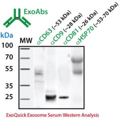
Exosome antibodies for Western blot analysis
For Western blotting analysis, we recommend resuspending the exosome pellet in 1XRIPA buffer with the appropriate protease inhibitor cocktail. SBI offers individual antibodies for CD63, CD9, CD81 and Hsp70 as well as a Western blot sampler kit (Catalog# EXOAB-KIT-1) which includes four exosomal marker antibodies: CD63, CD9, CD81, HSP70 (rabbit anti-human) and also includes a goat anti-Rabbit IgG HRP conjugated secondary antibody specificallytested for use in exosomal protein analysis. The primary antibodies are used at a 1:1,000 dilution and the HRP secondary antibody at 1:20,000 dilution.
Quantitate Exosomes by ELISA
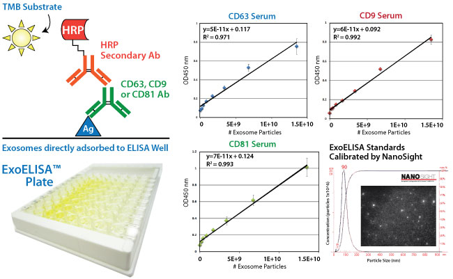
The ExoELISA kit is designed as a direct Enzyme-Linked ImmunoSorbent Assay (ELISA). The exosome particles and their proteins are directly immobilized onto the wells of the microtiter plate. After binding, wells are coated with a block agent to prevent non-specific binding of the detection antibody. The detection (primary) antibody is added to the wells for binding to specific antigen (e.g. CD63) protein on the exosomes.

A Horseradish Peroxidase enzyme (HRP) linked secondary antibody (goat anti-rabbit) is used for signal amplification and to increase assay sensitivity. A colorimetric substrate (Extra-sensitive TMB) is used for the assay read-out. The accumulation of the colored product is proportional to the specific antigen present in each well. The results are quantitated by a microtiter plate reader at 450 nm absorbance and calibrated by the exosome standards provided in the kit. The exosome standards are provided in number of exosome particles as determined by NanoSight dynamic light scattering measurements. The ExoELISA kits are compatible with exosomes isolated by ExoQuick, ExoQuick-TC or by ultracentrifugation techniques.
